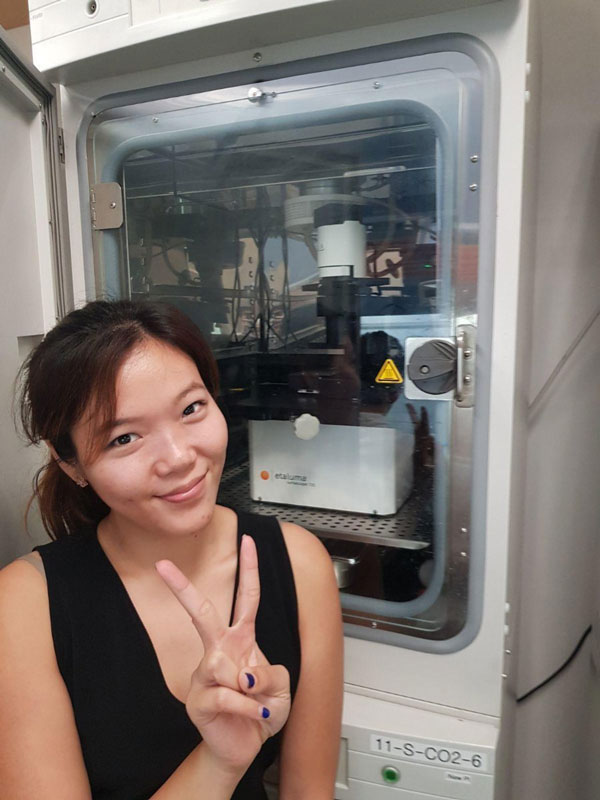
Crystal Chan with the Etaluma LS720 automated live cell microscope in an incubator at the Cancer Science Institute (CSI) Singapore - NUS
Crystal Chan with the Etaluma LS720 automated live cell microscope in an incubator at the Cancer Science Institute (CSI) Singapore - NUS
Crystal Chan with the Etaluma LS720 automated live cell microscope in an incubator at the Cancer Science Institute (CSI) Singapore - NUS
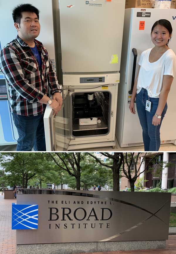
I really enjoyed my visit to the Mootha Lab at the Broad Institute yesterday. Big thanks to Tsz-Leung To and Wendy Hung who will be using our LS720 for single cell metabolomics studies.
I really enjoyed my visit to the Mootha Lab at the Broad Institute yesterday. Big thanks to Tsz-Leung To and Wendy Hung who will be using our LS720 for single cell metabolomics studies.
I really enjoyed my visit to the Mootha Lab at the Broad Institute yesterday. Big thanks to Tsz-Leung To and Wendy Hung who will be using our LS720 for single cell metabolomics studies.
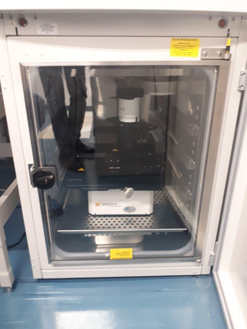
Congratulations to the Buxboim Lab in Israel for their recent installation of their new LS720. https://lnkd.in/dduNDSa
Congratulations to the Buxboim Lab in Israel for their recent installation of their new LS720. https://lnkd.in/dduNDSa
Congratulations to the Buxboim Lab in Israel for their recent installation of their new LS720. https://lnkd.in/dduNDSa
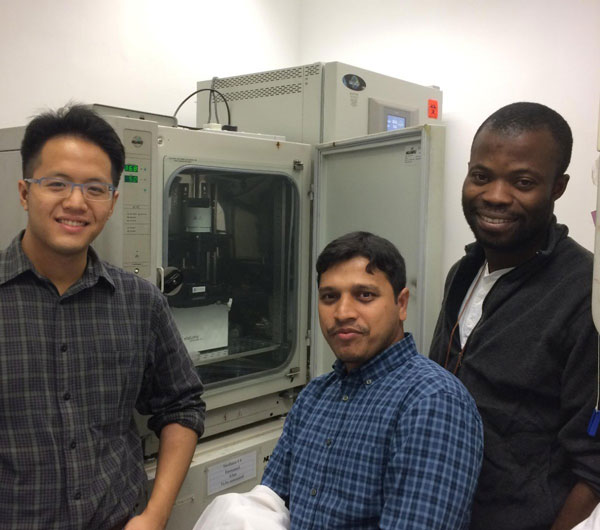
It was very nice to visit Dr. Amy Fong’s lab at City University of Hong Kong and meet Allan, Kowsar, and Emmanuel who are upgrading from their LS500 to the LS720 this year.
It was very nice to visit Dr. Amy Fong’s lab at City University of Hong Kong and meet Allan, Kowsar, and Emmanuel who are upgrading from their LS500 to the LS720 this year.
It was very nice to visit Dr. Amy Fong’s lab at City University of Hong Kong and meet Allan, Kowsar, and Emmanuel who are upgrading from their LS500 to the LS720 this year.
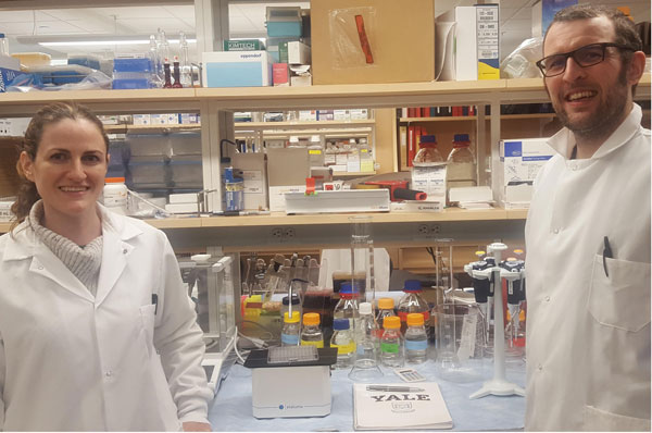
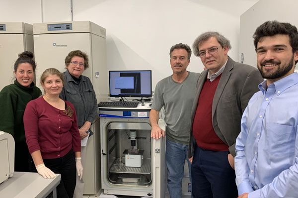
Assistant Professor Caroline H Johnson and Associate Research Scientist, Nicholas J. W. Rattray of the Environmental Health Sciences Department at the Yale School of Public Health, with the Etaluma, Inc. LS500 to be investigated for integration with mass spectrometry approaches.
Assistant Professor Caroline H Johnson and Associate Research Scientist, Nicholas J. W. Rattray of the Environmental Health Sciences Department at the Yale School of Public Health, with the Etaluma, Inc. LS500 to be investigated for integration with mass spectrometry approaches.
Assistant Professor Caroline H Johnson and Associate Research Scientist, Nicholas J. W. Rattray of the Environmental Health Sciences Department at the Yale School of Public Health, with the Etaluma, Inc. LS500 to be investigated for integration with mass spectrometry approaches.
I had a wonderful visit yesterday with Lori Roberts and James Maclean at Biotium. Look for an offering of simple live cell assays that run on our microscopes coming soon.
I had a wonderful visit yesterday with Lori Roberts and James Maclean at Biotium. Look for an offering of simple live cell assays that run on our microscopes coming soon.
I had a wonderful visit yesterday with Lori Roberts and James Maclean at Biotium. Look for an offering of simple live cell assays that run on our microscopes coming soon.
Big thanks to Kilean Lucas in Jim McGrath's lab too. Stay tuned for a VE-Cadherin timelapse in this neutrophil migration system featuring Etaluma's LS560 base scope with phase contrast by Olympus.
Big thanks to Kilean Lucas in Jim McGrath's lab too. Stay tuned for a VE-Cadherin timelapse in this neutrophil migration system featuring Etaluma's LS560 base scope with phase contrast by Olympus.
Big thanks to Kilean Lucas in Jim McGrath's lab too. Stay tuned for a VE-Cadherin timelapse in this neutrophil migration system featuring Etaluma's LS560 base scope with phase contrast by Olympus.
It's great to be working with Alec Salminen and Jim McGrath's group in Rochester. I'm looking forward to some revolutionary timelapse migration data very soon.
It's great to be working with Alec Salminen and Jim McGrath's group in Rochester. I'm looking forward to some revolutionary timelapse migration data very soon.
It's great to be working with Alec Salminen and Jim McGrath's group in Rochester. I'm looking forward to some revolutionary timelapse migration data very soon.
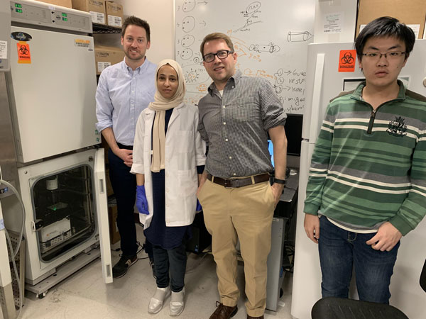
I had a great meeting with the Hartsough lab at Drexel University who is studying spheroid invasion into brain slices supported on transwell membranes. We are able to image through the membrane and tissue slice to watch the spheroid cells invade. Thanks to Ed Hartsough, Abeer Alharbi, Joshua Jackson, and Zongguan Huang.
I had a great meeting with the Hartsough lab at Drexel University who is studying spheroid invasion into brain slices supported on transwell membranes. We are able to image through the membrane and tissue slice to watch the spheroid cells invade. Thanks to Ed Hartsough, Abeer Alharbi, Joshua Jackson, and Zongguan Huang.
I had a great meeting with the Hartsough lab at Drexel University who is studying spheroid invasion into brain slices supported on transwell membranes. We are able to image through the membrane and tissue slice to watch the spheroid cells invade. Thanks to Ed Hartsough, Abeer Alharbi, Joshua Jackson, and Zongguan Huang.
This week we kicked off the winning research projects from the "Free Lumascope 620 for 6 Months" contest in the Fabrisia Ambrosio lab at UPMC International. Their two projects are entitled "Live cell imaging using the LS620 microscope to evaluate the impact of an “aged” substrate on muscle stem cell cytoskeletal and mitochondrial dysfunction" lead by Hikaru Mamiya and Amrita Sahu, and "Compressing Chronological Age by Means of Indirect Mixed Cell Cultures and Time-Lapse Imaging of Synthetic Exchange of Molecular Information in Muscle Stem Cells" lead by Amin Cheikhi. Here's a shot of abish pius, Sruthi Sivakumar, Hikaru Mamiya, and Amrita Sahu.
This week we kicked off the winning research projects from the "Free Lumascope 620 for 6 Months" contest in the Fabrisia Ambrosio lab at UPMC International. Their two projects are entitled "Live cell imaging using the LS620 microscope to evaluate the impact of an “aged” substrate on muscle stem cell cytoskeletal and mitochondrial dysfunction" lead by Hikaru Mamiya and Amrita Sahu, and "Compressing Chronological Age by Means of Indirect Mixed Cell Cultures and Time-Lapse Imaging of Synthetic Exchange of Molecular Information in Muscle Stem Cells" lead by Amin Cheikhi. Here's a shot of abish pius, Sruthi Sivakumar, Hikaru Mamiya, and Amrita Sahu.
This week we kicked off the winning research projects from the "Free Lumascope 620 for 6 Months" contest in the Fabrisia Ambrosio lab at UPMC International. Their two projects are entitled "Live cell imaging using the LS620 microscope to evaluate the impact of an “aged” substrate on muscle stem cell cytoskeletal and mitochondrial dysfunction" lead by Hikaru Mamiya and Amrita Sahu, and "Compressing Chronological Age by Means of Indirect Mixed Cell Cultures and Time-Lapse Imaging of Synthetic Exchange of Molecular Information in Muscle Stem Cells" lead by Amin Cheikhi. Here's a shot of abish pius, Sruthi Sivakumar, Hikaru Mamiya, and Amrita Sahu.
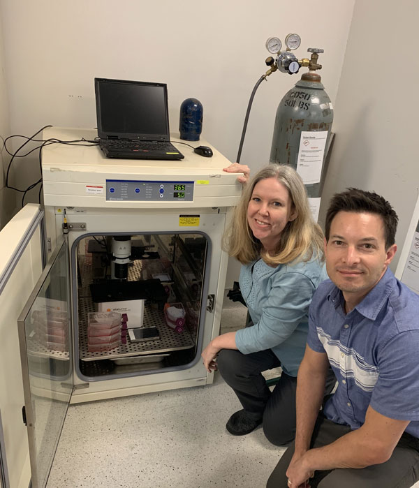
I enjoyed my visit to the D. Lansing Taylor labs at Pitt. Dillon Gavlock and Mark Miedel showed me the microfluidics imaging they are doing with oxygen gradients and primary hepatocytes on the Etaluma LS720.
I enjoyed my visit to the D. Lansing Taylor labs at Pitt. Dillon Gavlock and Mark Miedel showed me the microfluidics imaging they are doing with oxygen gradients and primary hepatocytes on the Etaluma LS720.
I enjoyed my visit to the D. Lansing Taylor labs at Pitt. Dillon Gavlock and Mark Miedel showed me the microfluidics imaging they are doing with oxygen gradients and primary hepatocytes on the Etaluma LS720.
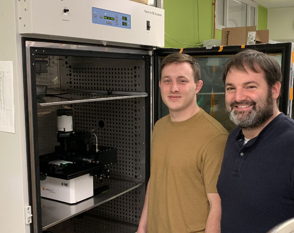
I had a great visit today with Dave Weaver at Vanderbilt University in Nashville where he put some very expensive Zeiss objectives on our LS620. Look for some great data from Dave's labs in the future!
I had a great visit today with Dave Weaver at Vanderbilt University in Nashville where he put some very expensive Zeiss objectives on our LS620. Look for some great data from Dave's labs in the future!
I had a great visit today with Dave Weaver at Vanderbilt University in Nashville where he put some very expensive Zeiss objectives on our LS620. Look for some great data from Dave's labs in the future!
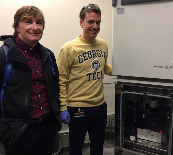
We had a nice visit with the Takayama microfluidics lab at Georgia Tech https://lnkd.in/evZNZzz. Matt Maudsley from Etaluma and Ph.D. candidate David R. Mertz pose next to their LS720 in an incubator.
We had a nice visit with the Takayama microfluidics lab at Georgia Tech https://lnkd.in/evZNZzz. Matt Maudsley from Etaluma and Ph.D. candidate David R. Mertz pose next to their LS720 in an incubator.
We had a nice visit with the Takayama microfluidics lab at Georgia Tech https://lnkd.in/evZNZzz. Matt Maudsley from Etaluma and Ph.D. candidate David R. Mertz pose next to their LS720 in an incubator.
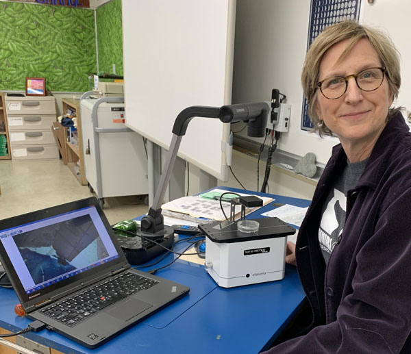
Etaluma is proud to donate a Lumascope to Julie Latta's class at Muirlands Middle School in La Jolla CA. Big thanks to Lamar Rutherford, MBA for recommending Julie. Looking forward to some cool microscopy images from the class.
Etaluma is proud to donate a Lumascope to Julie Latta's class at Muirlands Middle School in La Jolla CA. Big thanks to Lamar Rutherford, MBA for recommending Julie. Looking forward to some cool microscopy images from the class.
Etaluma is proud to donate a Lumascope to Julie Latta's class at Muirlands Middle School in La Jolla CA. Big thanks to Lamar Rutherford, MBA for recommending Julie. Looking forward to some cool microscopy images from the class.
I enjoyed working with our neighbors at SMSbiotech today. Big thanks to Camila, Maisson, Emily, Dr. Rahmo and Rob.
I enjoyed working with our neighbors at SMSbiotech today. Big thanks to Camila, Maisson, Emily, Dr. Rahmo and Rob.
I enjoyed working with our neighbors at SMSbiotech today. Big thanks to Camila, Maisson, Emily, Dr. Rahmo and Rob.
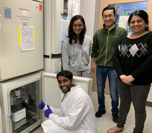
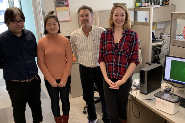
I had the pleasure of working with the Morris Lab at Washington University in St. Louis today. Big thanks to Kenji Kamimoto, Wenjun Kong, and Samantha Morris.
I had the pleasure of working with the Morris Lab at Washington University in St. Louis today. Big thanks to Kenji Kamimoto, Wenjun Kong, and Samantha Morris.
I had the pleasure of working with the Morris Lab at Washington University in St. Louis today. Big thanks to Kenji Kamimoto, Wenjun Kong, and Samantha Morris.
"Cell Tracking Analysis. Cells were seeded in 6-well plates and incubated for 24 h. LS620 Microscope (Lumascope) was used to record single cell position every 10min for 24h. Single cell migration distance and trajectory were analyzed by ImageJ."
"Cell Tracking Analysis. Cells were seeded in 6-well plates and incubated for 24 h. LS620 Microscope (Lumascope) was used to record single cell position every 10min for 24h. Single cell migration distance and trajectory were analyzed by ImageJ."
"Cell Tracking Analysis. Cells were seeded in 6-well plates and incubated for 24 h. LS620 Microscope (Lumascope) was used to record single cell position every 10min for 24h. Single cell migration distance and trajectory were analyzed by ImageJ."
I got to drop off a Lumascope to Eddie Adams, Ph.D. and our friends at Active Motif where Etaluma incubated in the early days.
I got to drop off a Lumascope to Eddie Adams, Ph.D. and our friends at Active Motif where Etaluma incubated in the early days.
I got to drop off a Lumascope to Eddie Adams, Ph.D. and our friends at Active Motif where Etaluma incubated in the early days.

We were thrilled to donate an LS500 microscope to Barb Phillips-Bredlow's 6th grade class at Northeast Nodaway School in Ravenwood, Missouri. Look for images on Twitter at @NEN6thgrade.
We were thrilled to donate an LS500 microscope to Barb Phillips-Bredlow's 6th grade class at Northeast Nodaway School in Ravenwood, Missouri. Look for images on Twitter at @NEN6thgrade.
We were thrilled to donate an LS500 microscope to Barb Phillips-Bredlow's 6th grade class at Northeast Nodaway School in Ravenwood, Missouri. Look for images on Twitter at @NEN6thgrade.

A great day at Newcastle University today with Julian Rutherford and colleagues imaging with 100x(oil). Looking forward to more tomorrow! Thanks Andrew Ridley
A great day at Newcastle University today with Julian Rutherford and colleagues imaging with 100x(oil). Looking forward to more tomorrow! Thanks Andrew Ridley
A great day at Newcastle University today with Julian Rutherford and colleagues imaging with 100x(oil). Looking forward to more tomorrow! Thanks Andrew Ridley
")
Thanks to Kirti Prakash for a great visit to the Rowitch Lab Wellcome Trust-MRC Cambridge Stem Cell Institute.
Thanks to Kirti Prakash for a great visit to the Rowitch Lab Wellcome Trust-MRC Cambridge Stem Cell Institute.
Thanks to Kirti Prakash for a great visit to the Rowitch Lab Wellcome Trust-MRC Cambridge Stem Cell Institute.

It was great working with Sally Wheatley, Kevin Gaston, and Morgan Thornber this week at the University of Nottingham.
It was great working with Sally Wheatley, Kevin Gaston, and Morgan Thornber this week at the University of Nottingham.
It was great working with Sally Wheatley, Kevin Gaston, and Morgan Thornber this week at the University of Nottingham.

A big thanks to Morgan Thornber at University of Nottingham for teaching Andrew Ridley and me about spider silk and bringing two specimens to donate silk for microscopy.
A big thanks to Morgan Thornber at University of Nottingham for teaching Andrew Ridley and me about spider silk and bringing two specimens to donate silk for microscopy.
A big thanks to Morgan Thornber at University of Nottingham for teaching Andrew Ridley and me about spider silk and bringing two specimens to donate silk for microscopy.

Testimonials
“We perform perfusion based live cell experiments in which we record the cell behavior while subjecting them to shear stress under flowing media conditions. In addition, we use the microscope to check transfection and transduction efficiency of fluorescently labeled proteins, which we like to combine with phase contrast bright field microscopy to see intra-cellular location of the expressed proteins. The ability to do perfusion imaging inside a cell culture incubator rather than within an acrylic enclosure on a traditional microscope is key. Keeping the reagents within the same incubator makes experimental design much easier and long-term experiments over days and weeks are much more stable. Flexible control software allows us to do the custom experiments we want. The small footprint of the instrument makes it very easy to transport from lab to lab and to integrate it in different experimental set-ups. Open access to the imaging labware for fluidics integration and a general open architecture allows us to bring in the other tools we want to the live cell imaging experiment. There is no maintenance required and the LEDs have an almost endless live-cycle. The instrument is super easy to handle and even unexperienced users are able to perform imaging after very little training (and we don’t need to be afraid that hardware breaks – robustness). As I see it now there are two camps. The expensive traditional confocal and now super resolution microscopes that push the borders of what technology can achieve and are mainly purchased by core centers. And the other is small and compact imaging systems that are affordable by small working groups and are designed for one or two of their specific applications. Take a look at all of your choices for microscopes beyond the big four and pay attention to hardware and software customizability. Especially customizability and the fast integration of additional software features is always much easier when working with smaller sized companies and start-ups, as communication is direct.”
“We have been using the Lumascope 600 fluorescent microscopes for about 5 years for teaching neurobiology and cell biology undergraduate lab courses and are very happy with them.
They are compact and portable, so we can move 8-10 scopes from lab to lab very easily, and they have been reliable and simple to use. We can get great image results for most of our experiments using a gain between 30-50% and a light intensity of 10-15%, thus maximizing image quality and minimizing potential problems from bleaching. The scopes are easy to use and stand up to the abuse that students can sometimes inflict and the scopes have held over the years with no technical malfunction issues or any wear being evident. The Lumaview software is both comprehensive and easy to use in terms of image acquisition and file storage and access.
At half the price of conventional upright fluorescent microscopes, and less than half the space taken up, the choice for us was clear and we have been more than satisfied. “
“I received the Lumascope and decided to set it up myself. Took it out of the box, installed the software, assembly and software were so intuitive that I didn’t even need the manual, and took this image in 15 minutes. Seriously. It is a prepared slide – but still … And I used my own high NA objectives!“
“Please let me take a moment to thank you for manufacturing such a wonderful microscope. Our Etaluma LS560 was simple to install and performed robustly right out of the box with easy to navigate software. But what is far more important is the insight this microscope provides us on the biological processes that are of greatest interest to us. In the past we have had to rely on static observations – which provide no access whatsoever to processes that are extremely dynamic; in our case changing dramatically minute by minute. We look forward to exploring these hidden worlds to better understand the biologies of our systems.”
“The Lumascope 720 has massively increased the amount of data that we can obtain from a single experiment, the ability to take images or videos of all wells on a single multiwell plate is amazing. My lab develops advanced image quantification analysis and we have been able to use all our previous pipelines (initially developed for images obtained on inverted epi’s or confocal) with almost no troubleshooting.”
“We had our first successful run yesterday on our new LS720. Everything ran fine according to plan – and at 4oC! We are impressed with the wide range of operating temperatures for the LS Microscopes and also the flexibility of integration. In fact, the additional TTL trigger on the LS720 enabled the easiest automation setup I have ever done. Great job!”
“Extraction of quantitative data from time-lapse imaging can provide unprecedented insights into cell signaling in single cells. A limiting factor in moving this field forward is the cost of existing live imaging systems, until now. The Lumascope 720 has enabled our small budget laboratory to extract real-time data from our multi-color live cell reporters. Thanks to the 720, we have been able to switch over from expensive ELISA based broken cell assays that measure the average signal across heterogeneous populations of cells at a single time point to now acquiring essentially free data points at single cell resolution over time. Also, thanks to the motorized stage and multiwell format we don’t need to use our plate reader anymore.”
“We have been working with Etaluma on developing the fluorescence imaging portion of our instrument. After looking at many other options, we selected a Lumascope optics module and are working with Etaluma to maximize our dye imaging signal and customize the module body. We have found this to be a very cooperative process, sharing ideas back and forth, with the common goal of providing a quality product that meets our specifications at an affordable price.”
“We love the Lumascope 500 because now we can get accurate transfection efficiencies!”
“…I wanted to tell you how pleased I am with the Lumascope…It is really easy to use and the students love it. As you know, I have worked in the fluorescence field for 40 years and I must say that if I had to choose three excitation wavelengths for a general use instrument such as yours, these are the wavelengths I would pick … you will hit the majority of the most commonly used probes … In fact, given the huge number of fluorophores commercially available, and the very wide range of spectroscopic capabilities they represent, in my opinion customers should be motivated to find fluorophores suitable for your instrument and appropriate for their research.
Congratulations for developing such a useful instrument!”
“The Etaluma allowed us to resolve low intensity fluorescent labeling that our existing Axiovert 200M could not.”
“The Lumascope is really [a] wonderful product!”
“Very easy to use. Students love it and results so far meet our expectations.”
“We really enjoyed your microscope and can see many potential uses in our lab. We are also very interested in the 720 and will consider looking into one of those later in the summer.”
“The Lumascope 620 provides far brighter and more uniform illumination than I have ever seen on any microscope using a standard Xe, Hg, or incandescent illuminator. Bravo! Etaluma microscopes are far more reliable than any standard microscope. LEDs are the most stable light source made. The illumination field is the most uniform I’ve seen. Digital calculations never change. Accuracy/precision seem to be within 3%, again superior to analog light sources or anything adjustable by hand. Our 620 scopes are in heavy use by students in several lab courses, as more instructors found out about them and saw how reliable they are, how easy to use, and how reasonably priced. I think they are even starting to creep into some research labs which don’t need the full facility of a Zeiss microscope. When I last looked, Etaluma was way ahead of the competition.”
Here are a few more of our happy customers.
Copyright © 2024 Etaluma, Inc. All rights reserved.